Better building, better science at Brooks Hall
July 22, 2026
New microscope could help unlock new ways to treat cancer
Bridget Plude watched amoeba cells crawl forward through a microscope. Then, she noticed little structures trailing out behind them and wondered what they were.
It was the start of a journey that led her to graduate school and a brand-new, powerful microscope, and into a field of inquiry that shows promise for new cancer treatments.
Plude, a graduate student in Central Michigan University’s biochemistry, cell and molecular biology program became one of just a small handful of people who’ve ever seen those little structures. They are called migrasomes because they form behind a cell as it migrates.
Migrasomes are a mystery to researchers, discovered only about a decade ago. Plude saw hers while studying a one-celled organism called Dictyostelium as an undergraduate in CMU’s biology program.
The promise of new discovery helped her decide to enroll in graduate school.
“I am totally in love with these cells and my research,” she said. “These cells I work with have become almost like my children. I work around their schedule, not mine. They have really opened my eyes to how privileged it is to work with a living organism every day.”
Dictyostelium are popular with researchers because they are simple and yet their genes are similar to those of people. Studying them helps scientists better understand basic biological and physiological processes in people.
Human cells also form migrasomes, for instance. Studying the relationship between Dictyostelium and its migrasomes can provide some illumination on the relationship between cells in the human body and their own migrasomes.
Certain cancer cells also form migrasomes, and that is where the possibility of future cures rests, she said. It starts with Dictyostelium’s version of an empty stomach.
Dictyostelium normally live as independent amoebas in the soil, consuming bacteria. When food becomes scarce, they come together to form a single unit that creates a stalk to distribute spores so that some of the amoebas find a new home.
The behavior mirrors that of certain metastasizing cancer cells, Plude said. Migrasomes appear to play a role, but additional research is required because migrasomes themselves are relatively new discoveries.
“Ten years is nothing in terms of how much knowledge we really need to understand something as a unit,” she said.
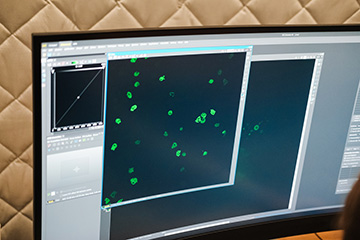
At first glance, migrasomes look a bit like pomegranates, with an outer membrane holding in individual pouches, she said. It’s believed that the pouches contain different structures that serve multiple purposes.
Migrating cells pull the migrasome behind them with a tether. It’s believed that the tethers serve as a conduit between the cell and migrasome, relaying information and possibly waste products from inside the cell, Plude said.
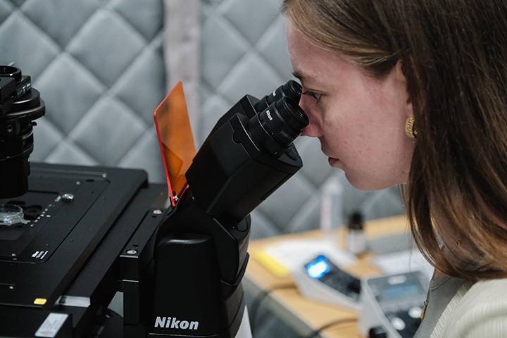
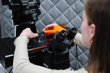
Looking inside the pouches to see what they contain would answer what cells use migrasomes for. Plude said she’s seen the pouches. She hasn’t seen what is in the pouches, however. The microscope she first saw them through wasn’t powerful enough to get that detailed.
A timely equipment upgrade will help her finally do that.
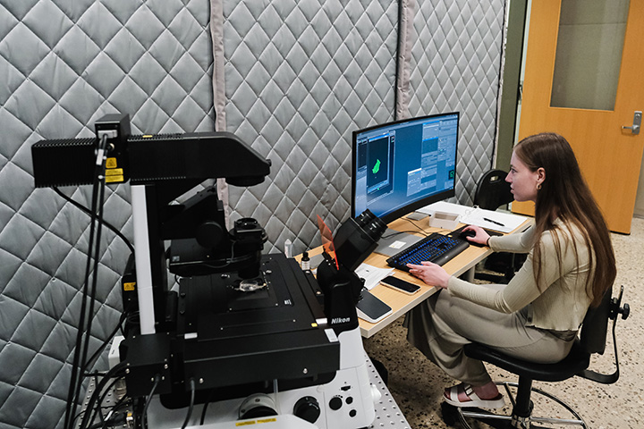
In May, CMU installed a new $730,000 microscope with advanced imaging abilities that can turn a stack of two-dimensional images into a three-dimensional model. Its magnification level also allows users to put something the size of a cell onto a viewing screen.
Plude is already using it. Once she’s more comfortable using it, she said she hopes to get a close look into what the pouches contain. That could confirm that migrasomes serve multiple functions, from cell-to-cell communication to waste disposal.
Cells need to get rid of waste like degraded mitochondria, which generate energy for the cell, and replace them with new ones. Plude would like to know if Dictyostelium discards used-up mitochondria by moving them to migrasomes via the protein tether.
She said confirming that would open up research into whether blocking cells from moving spent mitochondria to the migrasomes could kill them. If it works in cancer cells, it is theorized that it could prevent cancer from spreading.
There is already hopeful research involving several different cancers, including a kind of brain cancer, a kind of liver cancer, a kind of bone cancer and a kind of breast cancer.