NIH grant allows biosciences lab to break new ground with cutting-edge microscope
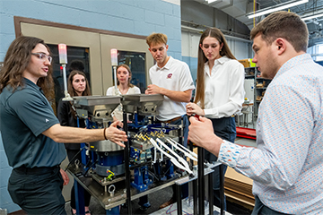
In a groundbreaking move, Jennifer Schisa’s Biosciences laboratory has propelled itself into the forefront of scientific discovery with the acquisition of a specialized Zeiss Apotome 3 microscope system, made possible by a generous $100,000 supplemental grant from the National Institutes of Health. The installation of this state-of-the-art fluorescence microscope marks the beginning of an exciting chapter, as researchers and students eagerly embark on a journey into uncharted territories of cellular structures and protein dynamics.
This cutting-edge technology not only expands the scope of visualizable fluorescently-tagged proteins but also boasts high-resolution imaging capabilities comparable to confocal microscopes. What sets it apart is its user-friendly design, making it accessible to students who are actively engaged in data collection to finalize a groundbreaking publication.
The newfound capabilities of the microscope have revolutionized the possibilities within the lab, enabling students to undertake experiments that were previously impossible.
The advanced technology has opened the door to answering new questions as researchers explore the intricate world of proteins, unhampered by previous technological constraints.
Undergraduate researcher Christya Haddad is at the forefront of these groundbreaking experiments, investigating changes in the actin cytoskeleton under conditions where unusual protein granules form in eggs. The most effective fluorescent tag on actin, visible only at specific wavelengths, can now be used in the lab, providing crucial insights into the structural alterations triggering the formation of protein granules.
Ashley Cichon, another undergraduate, is shedding light on the formation of unusual granules in eggs, focusing on the Conserved Germline Helicase-1 protein. The Zeiss Apotome 3 microscope allows her to visualize this key protein for the first time in the lab, unraveling intricate processes within the eggs.
Graduate student Victoria Tice is delving into the consequences of unusual protein granules in eggs. Preliminary results indicate at least one gene in developing eggs is expressed earlier than usual when granules form, supporting the hypothesis that such granules may lead to low-quality eggs.
In a collaborative effort, a team of graduate and undergraduate students is embarking on a project to identify novel regulators of protein granules in eggs. Leveraging the microscope's high resolution, they aim to efficiently examine approximately 100 genes within a few months, seeking to determine the genes influencing the formation of protein granules.
These experiments hold implications beyond academic curiosity. Researchers aspire to understand if the formation of unusual protein granules indeed results in low-quality eggs, with the ultimate goal of paving the way for new therapeutic interventions to enhance fertility in women. As the studies progress, the researchers plan to compile their findings into a manuscript for submission in the coming months, anticipating that their discoveries may shape the future of reproductive biology.