Now through October 31, we're waiving application fees for all freshman and transfer applicants!
*Common app code: Central27
Student Stories Discovery in Focus
Your experience at CMU can take you places you never imagined. Through hands-on learning and access to advanced microscopy and imaging technology, you’ll build skills, explore new questions, and discover opportunities that extend beyond the classroom. Hear from current and former students as they share how their experiences helped shape their academic journeys, inspire their research, and prepare them for what comes next. Their stories offer a glimpse of what’s possible when you turn your curiosity into discovery.
Abby (Tirrell) Vandenberg
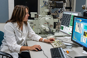
Abby (Tirrell) Vanderberg graduated from the CMU microscopy program in 2008. In 2009 she started working as a Scanning Electron Microscopist at Michigan State University’s Center for Advanced Microscopy, where she still works today. The Center for Advanced Microscopy is a core facility that supports over 40 different departments at MSU. Abby’s role includes training graduate students in the use of scanning electron microscopes and collaborating with researchers on a wide range of samples, from plants and microbes to polymers and nanoparticles.
Joseph Harris
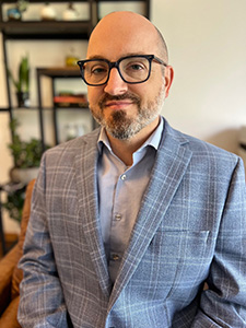
Joseph Harris received his BS in Biology: Microscopy from Central Michigan University in 1999; since 2000 he has worked as a Technologist in Dow Chemical's Analytical Sciences division of Core Research and Development. Joe has over 25 years of experience in microscopy, particularly TEM and SEM. He uses advanced microscopy methods to analyze polymeric materials across all Dow businesses, contributing to every part of the innovation cycle. In his current role, he is responsible for maintaining instruments, training colleagues and working with business partners to contribute toward product development. His research of halogen free flame retardants, nanocomposites and polyurea grease have been published in major scientific journals. He has developed innovative sample prep and staining methods to understand the fundamental structure-property relationship in polyolefin dispersions for can and paper coatings. These contributions helped Dow launch two new products (CANVERA™ 1110 and RHOBARR™ 320) and earn the team two R&D 100 awards, an Edison gold award and Lux award for sustainability. Joe believes that the early hands-on experience and training he received at CMU were crucial in laying the groundwork for his successful microscopy career.
Alicia Trombley
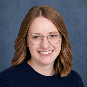
“Pursuing a microscopy concentration proved foundational to both my academic path and my current career. The undergraduate research requirement led me to join a lab where I gained hands-on research experience and ultimately remained to complete a master's degree in biology. My thesis relied heavily on microscopy techniques, strengthening my technical expertise and understanding of experimental design. I later built on this scientific background by earning a law degree at Michigan State University where I focused on intellectual property law. Today, I combine my research experience and legal knowledge in my role as a Research Compliance Specialist Lead for the University of Michigan.” - Alicia Trombley (graduated with B.S. in 2014 and M.S. in 2016)
Kevin Schmidt
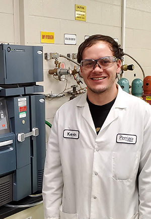
My name is Kevin Schmidt, and I graduated from CMU in 2015 with a major in Biology and a concentration in Microscopy. I joined Perrigo in March 2016 and currently work there as an MRB (Material Review Board) Associate Scientist. In this role, I collaborate with a cross-functional team to evaluate supplier changes and proposed alternate source materials, assessing their potential impact on our commercial drug products. The Microscopy program equipped me with a broad set of laboratory skills and techniques that were able to transfer over to my current career path.
Debbie Rothe
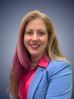 “Vision is the art of seeing what is invisible to others.” — Jonathon Swift.
“As a microscopist, I appreciate how much we learn by making things visible that would go otherwise unseen with the unaided eye. I’m grateful for the chance I had as a CMU undergraduate to enroll in microscopy courses, which sharpened my technical abilities and helped me connect with fellow microscopists throughout Michigan. These experiences ultimately paved the way for a rewarding career at Dow.
I still recall being granted permission to take SEM and TEM classes during my undergrad years. I learned valuable lessons about meticulous sample preparation and the attention to detail required for producing quality images. I also learned to start by considering the broader context before focusing on smaller areas of interest.
This foundation led me to a co-op position with Dow’s Microscopy Group in R&D Analytical Sciences, where I worked alongside others who were equally passionate about microscopy and problem-solving. My success there resulted in an offer for a full-time position upon earning my bachelor’s degree.
My journey began in the microscopy darkroom, prior to Dow’s shift to digital imaging, where I handled both sample prep and some analysis. Over time, I expanded my skills into microanalytical techniques, becoming a valued problem solver for Dow’s products and manufacturing operations. While capturing appealing images was important, the real value lay in accurately interpreting results for clients and communicating those findings to non-specialists. This enabled better decisions for product development and manufacturing.
As my passion for materials science and solving problems deepened, I returned to school to pursue a Ph.D., further expanding my expertise and leadership potential. This advancement allowed me to lead larger projects and make a greater impact. I have guided teams including analysts, environmental data experts, and waste specialists. Currently, I lead a team of environmental experts who are responsible for delivering sustainable waste solutions that benefit both Dow and the planet. I also serve as Change Lead for Dow’s Global Water & Nature Strategy, where I develop and execute plans to increase organizational awareness of water and habitat conservation opportunities, supporting Dow in reaching its public water and nature goals.
What began in the basement of Brooks Hall became the cornerstone of a multi-decade career at a Fortune 100 company. I remain deeply thankful to the professors and staff who shaped my journey and am delighted to see this program thriving at my alma mater!” – Debbie Rothe
Joe Patterson
- B.S. Biology (Microscopy), Central Michigan University 2008
- M.S. Biology, Central Michigan University 2010
- Ph.D. Genetics, Michigan State University 2016
Lab Webpage: https://translationalscience.msu.edu/research-groups/patterson-lab.html
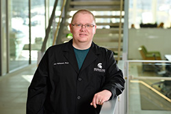
“I am currently an assistant professor in the Translational Neuroscience department at Michigan State University (Grand Rapids) where my lab focuses on the impact of comorbidities on the onset/progression of neurodegenerative diseases such as Parkinson’s disease. My lab regularly uses light microscopy (simple stains, immunohistochemistry, immunofluorescence, stereology, etc.) and transmission electron microscopy (negative staining of protein fibrils to assess size and structure) in our projects.
Elements from the microscopy program at CMU have been foundational to my career both in graduate school and in my current position. The opportunity to develop projects based on each microscope, learn sample preparation, and troubleshoot methods prepared me for lab work in graduate school. The focus on independence in the 403 undergraduate research project and class projects established my foundation on how to synthesize and apply information from the literature and reference texts, which is critical to my current position.”
Cooper Allers
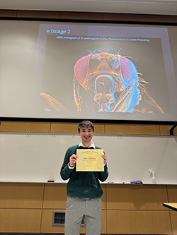 “Being a part of the microscopy program was a very fulfilling experience for me. It was always something that I had been passionate about, and the microscopy program allowed me to engage with my interests. In Spring of 2023, I took the scanning electron microscopy course, and I loved it. I loved the attention to detail that it demanded and the feeling of getting a good image. From that class, I was able to join Dr. Steinhilb's lab working with Drosophila. I imaged drug treated flies that expressed an Alzheimer's protein, and our goal was to identify treatments that qualitatively lowered Alzheimer's presentation in this model organism. Through that experience, I got to take some great images, and I really enjoyed honing the craft. The classes that I took and the work that I did confirmed for me that I wanted to go to medical school to become a pathologist in a clinical lab. In Spring of 2025, I graduated from CMU, and in August, I continued onto medical school at CMU. In my time here so far, the skills that I learned in the microscopy program have helped me immensely in my learning and understanding of medicine. My biggest takeaway from the program is that microscopy is truly an art, and that it takes care, time, and dedication to be good at it.” – Cooper Allers
Cody Morrison
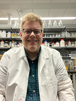
“I am currently a fourth-year PhD Candidate in Dr. Cynthia Damer’s lab. My research focuses on investigating the cellular function of copine proteins in the model organism Dictyostelium discoideum. I will be graduating in August of 2026 and anticipate joining a research lab somewhere as a postdoctoral fellow. In the future, I hope to run my own lab at a university as a principal investigator and build a career uncovering the role autophagy plays in cancer or neurodegeneration. The microscopy facilities, along with the training I have received from faculty and staff, have taught me how to troubleshoot experimental design issues, as well as how to efficiently display results to any audience.” – Cody Morrison
Devon Leroux
Degree:
- B.S., Biology; Microscopy Concentration
- M.S., Biology
Title: Electron Microscopy Sample Prep Specialist – Thomas Scientific
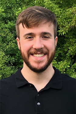
“CMU’s Microscopy program provided me with unique opportunities that opened doors in my academic and professional careers. As an undergraduate, students develop hands-on microscopy skills with niche systems in the fields of confocal, SEM, and TEM. Small class sizes and experienced professors help students flourish. As an undergraduate, this experience allowed me to play a meaningful role in active research and prepared me for my eventual master’s program at CMU. After completing my graduate degree, I was hired directly into the University of Michigan due to the breadth of microscopy skills that I had gained as a student. At UM, I worked as an EM Specialist supporting researchers across the university. It was in this role that I met my current employer and transitioned to selling and supporting the very EM instrumentation that I learned to operate at CMU. The microscopy program laid the foundation for my career and many others.”
Ty Nelson
“Being a part of the microscopy program at CMU has been a great learning experience. What I enjoy the most about being in this program is the hands-on aspect of the courses. Each course I’ve taken in the program teaches you the necessary skills needed to perform various methods of microscopy. The techniques of preparing samples and operation of microscopes can seem intimidating and challenging at first, but I found that practicing and doing it more frequently made it less difficult. During my time in the program, I’ve learned to use light microscopes, confocal microscopes, and electron microscopes. Using the different microscopes was interesting to me because it allows us to view things ordinarily not seen.
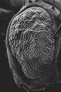
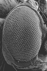
During the 2025 Spring semester I joined Dr. Steinhilb’s research lab to take images of Drosophila melanogaster using the scanning electron microscope. Certain genotypes of these fruit flies show what is called “tau toxicity” in their eyes. This toxicity is caused by damaged eye neurons due to abnormally phosphorylated tau proteins. These abnormal tau proteins also play a crucial role in the development of Alzheimer’s and other neurodegenerative diseases. The goal of the research lab is to see if there’s a drug that can prevent these tau proteins from damaging the flies’ eyes to forward Alzheimer’s research. The images I took were used to assess whether the drugs were working based on the condition of the flies’ eyes.” – Ty Nelson
Brie Plude
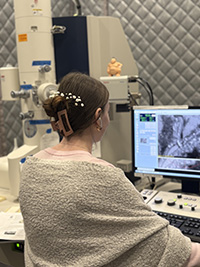
"My name is Brie Plude, and I am attending Central Michigan University in hopes of obtaining a PhD in Biochemistry, Cell, and Molecular Biology. My work focuses on understanding how proteins, called copines, play a role in cell migration and cell–cell communication. To investigate these processes, I am able to use several advanced imaging techniques available through the university’s imaging facility: confocal microscopy to examine protein localization and dynamics during migration, Total Internal Reflection Fluorescence (TIRF) microscopy to visualize protein localization events occurring at or near the plasma membrane, and Transmission Electron Microscopy (TEM) to examine cellular structures at micron-level resolution. I have made numerous discoveries within CMU's imaging facility, and it has been so much fun!"