Teeth: Sign of Stress
 Anthropology professor Dr. Cathy Willermet developed a non-destructive method for analyzing defects of dental enamel (DDE). Dental enamel is the hardest substance in the human body and protects teeth from most chemical or thermal substances that may harm the inner tooth. Since dental enamel preserves well, it is often used for studying archeological human remains. Using specialized microscopes, Dr. Willermet and her co-authors studied the factors that influence DDE, with a focus on stress.
Many bioarchaeologists are interested in determining a connection between early life stress and mortality. Since dental enamel develops as teeth do, researchers can gain valuable insight into a subject’s early life by analyzing the linear enamel hypoplasia (LEH) and striae of Retzius found in tooth enamel. LEH are lines of reduced enamel that appear on the surface of a tooth and result from stress. Striae of Retzius are growth lines in enamel that can help give information about the frequency of the stress.
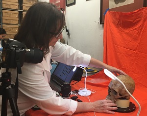
Dr. Willermet developed a cost-efficient and non-destructive method for studying developmental stress in teeth: digital light microscopy (DLM). DLM combines the technology of a traditional microscope with the imaging component of a digital camera. With this technology, the research team photographed human canine teeth (collected from the Maxwell Museum Documented Skeletal Collection) and analyzed the DDE by looking at the spacing between the striae of Retzius lines. While there are many methods used for examining tooth enamel they can be expensive, destructive, and tedious, DLM is non-destructive, cost-efficient and relatively easy.
This new method can be applied to large samples of teeth and contribute to the growing understanding of DDE and developmental stress in populations.
At CMU We Do Research, We Do Real World
Story by ORGS intern Hailey Nelson
December 2021